The 64-slice MDCT allows diagnostic and interventional medicine to merge. Fast, accurate imaging tools can advance medicine so that the diagnosis and treatment can occur on the same day. Integrated high resolution 3D imaging with more complex image guidance systems can, eventually, increase the effectiveness of the initial procedure and eliminate follow-up ablation procedures. A few systems are in development including:
 A robot arm with a gamma sensor controlled by the 3D coordinate system of the MDCT. The gamma sensor detects molecular imaging contrast material which may be used to illuminate denatured proteins or cell necrosis. [3]
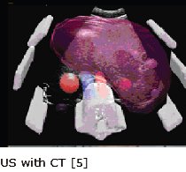
- Live 3D ultrasound overlaid on a retrospective CT image can improve visualization during targeting when gas, formed from the burn, or bone create shadows on the CT. [5] The image on the right shows a real time US of three simulated markers merged with segmented CT of a test model.
When integrated with the real time 3D coordinate system of the MDCT, these techniques can improve visualization and monitoring of the treatment, increase the likelihood of successful tumor ablation, and improve the patient outcome.